
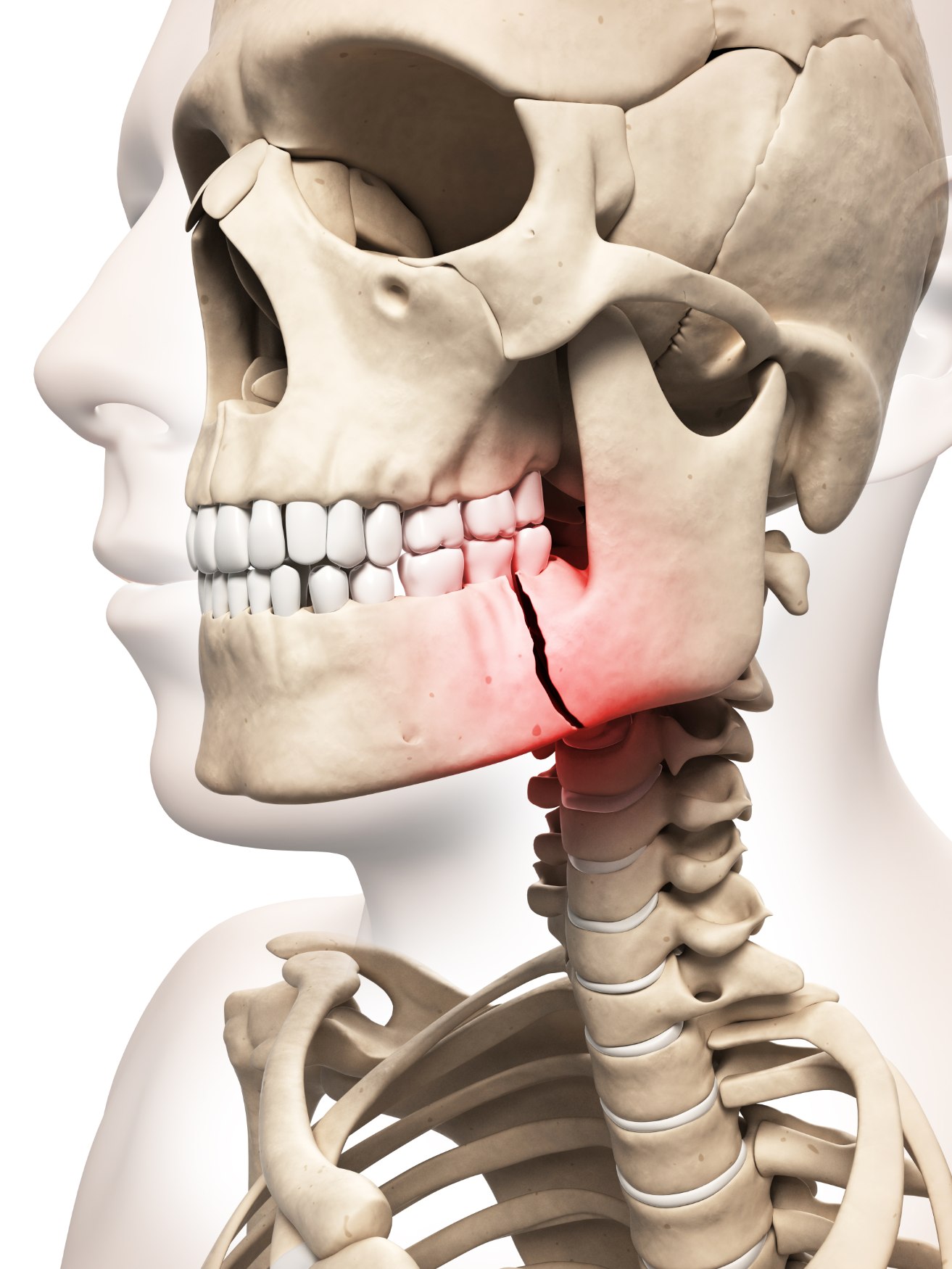
Złamanie szczęki i żuchwy to urazy, do których dojść może na wiele różnych sposobów.
Niezależnie od okoliczności powstania urazu złamania szczęki i żuchwy wymagają
odpowiedniego leczenia podjętego przez chirurga. Tylko w ten sposób możemy odzyskać
dawną funkcjonalność narządu żucia i komfort życia. Niektóre złamania szczęki nie są
widoczne na pierwszy rzut oka i można ich nawet nie odczuwać. Mogą one jednak
prowadzić do wady zgryzu, dlatego często są one wykrywane podczas wizyty u dentysty.
Ponadto, nieleczone złamanie szczęki może skutkować trwającą całe życie deformacją
twarzy, która staje się bardzo trudna do wyleczenia na późniejszym etapie.
Nasi specjaliści wykorzystują najnowszą diagnostykę, taką jak CBCT, tomografia
komputerowa, skan Panorex, aby zidentyfikować wszystkie złamania i opracować
odpowiedni plan leczenia. Wykorzystują również najnowocześniejszą technologię druku 3D
do rekonstrukcji wtórnych ubytków urazowych, które nie są podatne na tradycyjne metody
leczenia.
Facial fractures are unique in a way – if the bones are not aligned and fixed correctly, they usually lead to the deranged bite or a malocclusion. Depending on the location of the break or fracture, and the severity of the injury, your surgeon may offer a number of different treatment options. Duration of treatment can differ significantly depending on the individual condition, and in cases where there is only a minor fracture, effective management of the injury is still essential. Some jaw fractures have slight or no displacement, and the individual may look uninjured; however, the complaint of a subtle malocclusion leads their dentist to suspect a fracture.
In addition, an untreated jaw fracture can also result in lifelong facial deformity, which becomes very difficult to treat at a later stage. Our specialists use the latest diagnostic modalities like CBCT, CT scan, and Panorex scan to identify all the fractures and formulate a suitable treatment plan. They also use state-of-the-art 3D printing technology for the reconstruction of secondary traumatic defects, which are not susceptible to traditional treatment options.
In addition, an untreated jaw fracture can also result in lifelong facial deformity, which becomes very difficult to treat at a later stage. Our specialists use the latest diagnostic modalities like CBCT, CT scan, and Panorex scan to identify all the fractures and formulate a suitable treatment plan. They also use state-of-the-art 3D printing technology for the reconstruction of secondary traumatic defects, which are not susceptible to traditional treatment options.
Procedure
Sometimes, elastic bands may be used to guide the jaw into position following surgery. These are attached using small temporary metal wires or braces. Your procedure will be discussed with you in full during your consultation. Even the fractured zygomaticomaxillary complex can be managed and often treated from an intraoral approach, anatomically reduced and fixated in the presence of gross facial oedema. The days of the ‘Gillies’ lift technique are being replaced by the intra-oral buttress or other methods with rigid fixation. These facial fractures are often considered a simple treatment for Oral and Maxillofacial surgeons, as it is simply a matter of putting the puzzle pieces back together.