
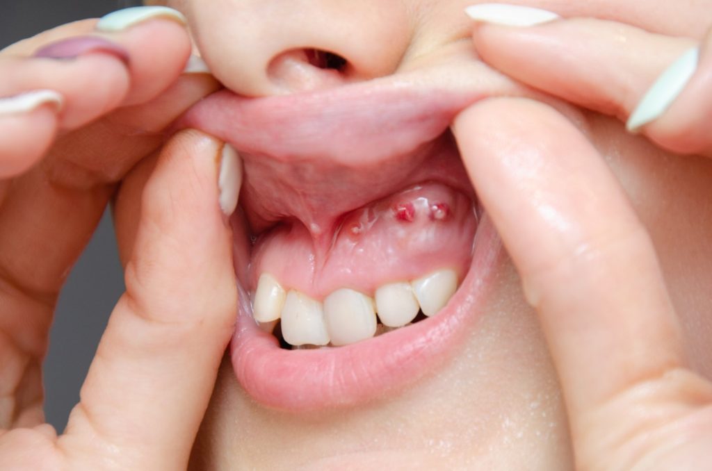
Les lésions kystiques des mâchoires sont des changements
pathologiques courants. Ce groupe de lésions comprend de
nombreuses affections, qu’elles soient odontogènes ou non
odontogènes. Les kystes odontogènes proviennent des restes de tissus
de formation dentaire et, par conséquent, ils se développent dans les
zones dentées des mâchoires. On pensait classiquement que les kystes
nodontogènes provenaient des restes des processus maxillaires ou
mandibulaires dans l’embryon en formation. De nombreux kystes sont
à croissance lente et asymptomatiques, souvent trouvés comme des
découvertes fortuites lors des examens dentaires. Parfois, les kystes
provoquent la migration des dents, ce qui entraîne un espacement ou
un encombrement entre les dents. Dans de rares cas, si les kystes ne
sont pas traités, ils peuvent provoquer une érosion excessive des os de
la mâchoire, entraînant des fractures pathologiques de la mâchoire. Un
diagnostic précis et un traitement approprié sont nécessaires, car les
lésions continuent généralement de croître et peuvent affecter les dents
adjacentes et d’autres structures ou même entraîner une fracture
pathologique. Une fois qu’un kyste est suspecté, nos spécialistes
conseillent des radiographies et des scintigraphies CBCT pour définir
l’étendue de la lésion et formuler un plan de traitement en
conséquence. Dans certains cas, les médecins vérifient le kystique et
l’envoient pour un examen microscopique qui donne un diagnostic sur
la nature du kyste. Certains kystes sont petits et peuvent être enlevés
par le dentiste sous anesthésie locale. Cependant, la plupart des kystes
nécessitent un traitement chirurgical plus large. Les tumeurs
odontogènes, par définition, sont dérivées de tissus dentaires et, bien
qu’elles aient été signalées dans des structures non dentaires, elles se
développent principalement dans le maxillaire ou la mandibule. La
majorité des tumeurs sont bénignes et, bien qu’elles puissent être
localement agressives, elles peuvent rarement métastaser. Les tumeurs
nodontogènes de la mâchoire, quant à elles, se développent à partir
d’une grande variété de tissus du corps, proviennent souvent des os
faciaux qui ne portent pas de dents et peuvent se développer dans
d’autres endroits à l’extérieur de la tête et du cou. Si les tumeurs sont
laissées sans traitement, elles peuvent atteindre une taille
disproportionnée et causer l’affaiblissement des os de la mâchoire,
conduisant éventuellement à la fracture pathologique de l’os. Ils sont
également plus susceptibles d’être infectés, ce qui complique
davantage le traitement. Les médecins commenceront par un examen
clinique, une biopsie et des radiographies, une TDM ou une CBCT
pour identifier la nature de la tumeur. Le traitement est déterminé par
Cystic lesions of the jaws are common pathologic changes. This group of lesions includes many conditions, either odontogenic or nonodontogenic. Odontogenic cysts arise from the remnants of tooth formation tissues, and consequently, they develop in the tooth-bearing areas of the jaws. Nonodontogenic cysts were classically thought to arise from the remnants of the maxillary or mandibular processes in the forming embryo. Many cysts are slow growing and asymptomatic, often found as incidental findings at dental check-ups. Sometimes cysts cause the migration of teeth, resulting in spacing or crowding between the teeth.
In rare cases, if cysts are left untreated, they can cause excessive erosion of the jaw bones, resulting in pathologic fractures of the jaw. Accurate diagnosis and appropriate treatment are required, as lesions generally continue to grow and may affect adjacent teeth and other structures or even result in pathologic fracture. Once a cyst is suspected, our specialists advise x-rays and CBCT scans to define the extent of the lesion and formulate a treatment plan accordingly. In some cases, doctors check the cystic and send it for a microscopic examination which gives a diagnosis about the nature of the cyst. Some cysts are small and can be removed by the dentist with local anaesthesia. However, most cysts require wider surgical treatment. Odontogenic tumours, by definition, are derived from tooth-related tissues, and although they have been reported in non-tooth-bearing structures, they develop mainly in the maxilla or mandible. The majority of tumours are benign, and while they may be locally aggressive, they rarely can metastasise. Nonodontogenic jaw tumours, on the other hand, develop from a wide variety of tissues in the body, often originate in non-tooth-bearing facial bones, and may develop in other sites outside of the head and neck. If tumours are left without treatment, they can grow to a disproportionate size and cause the weakening of the jaw bones, eventually leading to the pathologic fracture of the bone. They also are more prone to get infected, which further complicates the treatment. Doctors will begin with clinical examination, biopsy and x-rays, CT or CBCT scans to identify the nature of the tumour. Treatment is decided by numerous factors like – age, side, nature and size of the tumour. The management of these pathological changes is not just limited to removal but also involves the rehabilitation of the jaw bones. Since jaw bones, unlike other bones in the human body, give form and shape to our facial structure, it becomes necessary to plan their reconstruction. While most of the tumours are small and can be appropriately managed by resection and rehabilitation by dental prosthesis, some tumours involve the entire height of the bone, which require segmental resection of the jaw bone. This requires meticulous planning, for which specialists use 3-D and virtual surgical planning software to identify the exact amount of bone to be resected and decide about the reconstruction modality. Defects can be reconstructed by taking autogenous bones or bones from other parts of the same individual. However, some defects require reconstruction with a prosthesis, which is a 3D facial implant that mimics the natural bone contour, thereby restoring jaw continuity.