
كسور الوجھ فریدة من نوعھا بطریقة ما – إذا لم یتم محاذاة العظام وتثبیتھا بشكل
صحیح ، فإنھا عادة ما تؤدي إلى العضة المختلة أو سوء الإطباق. اعتمادا على موقع الكسر أو الكسر
، وشدة الإصابة ، قد یقدم الجراح عددا من خیارات العلاج المختلفة. یمكن أن تختلف مدة العلاج
اختلافا كبیرا اعتمادا على الحالة الفردیة ، وفي الحالات التي لا یوجد فیھا سوى كسر بسیط ، لا تزال
الإدارة الفعالة للإصابة ضروریة. بعض كسور الفك لھا إزاحة طفیفة أو معدومة ، وقد یبدو الفرد
غیر مصاب. ومع ذلك ، فإن الشكوى من سوء الإطباق الدقیق تقود طبیب الأسنان إلى الاشتباه في
حدوث كسر. بالإضافة إلى ذلك ، یمكن أن یؤدي كسر الفك غیر المعالج أیضا إلى تشوه في الوجھ
مدى الحیاة ، والذي یصبح من الصعب جدا علاجھ في مرحلة لاحقة. یستخدم المتخصصون لدینا
أحدث طرق التشخیص مثل الكسور. كما أنھا تستخدم أحدث تقنیات الطباعة لإعادة بناء العیوب
المؤلمة الثانویة وصیاغة خطة علاج مناسبة ، والتي لیست عرضة لخیارات العلاج التقلیدیة.
الإجراء: في بعض الأحیان ، یمكن استخدام الأربطة المرنة لتوجیھ الفك إلى موضعھ بعد الجراحة.
یتم إرفاق ھذه باستخدام أسلاك معدنیة صغیرة مؤقتة أو الأقواس. ستتم مناقشة الإجراء الخاص بك
معك بالكامل أثناء استشارتك. حتى المركب الوجني الفكي المكسور یمكن إدارتھ وغالبا ما یتم علاجھ
من خلال نھج داخل الفم ، ویتم تقلیلھ تشریحیا وتثبیتھ في وجود وذمة الوجھ الإجمالیة. یتم استبدال
تقنیة الرفع بدعامة داخل الفم أو طرق أخرى ذات تثبیت صلب. غالبا ما تعتبر كسور الوجھ ھذه
.علاجا بسیطا لجراحي الفم والوجھ والفكین ، حیث إنھا ببساطة مسألة إعادة تجمیع قطع اللغز معا
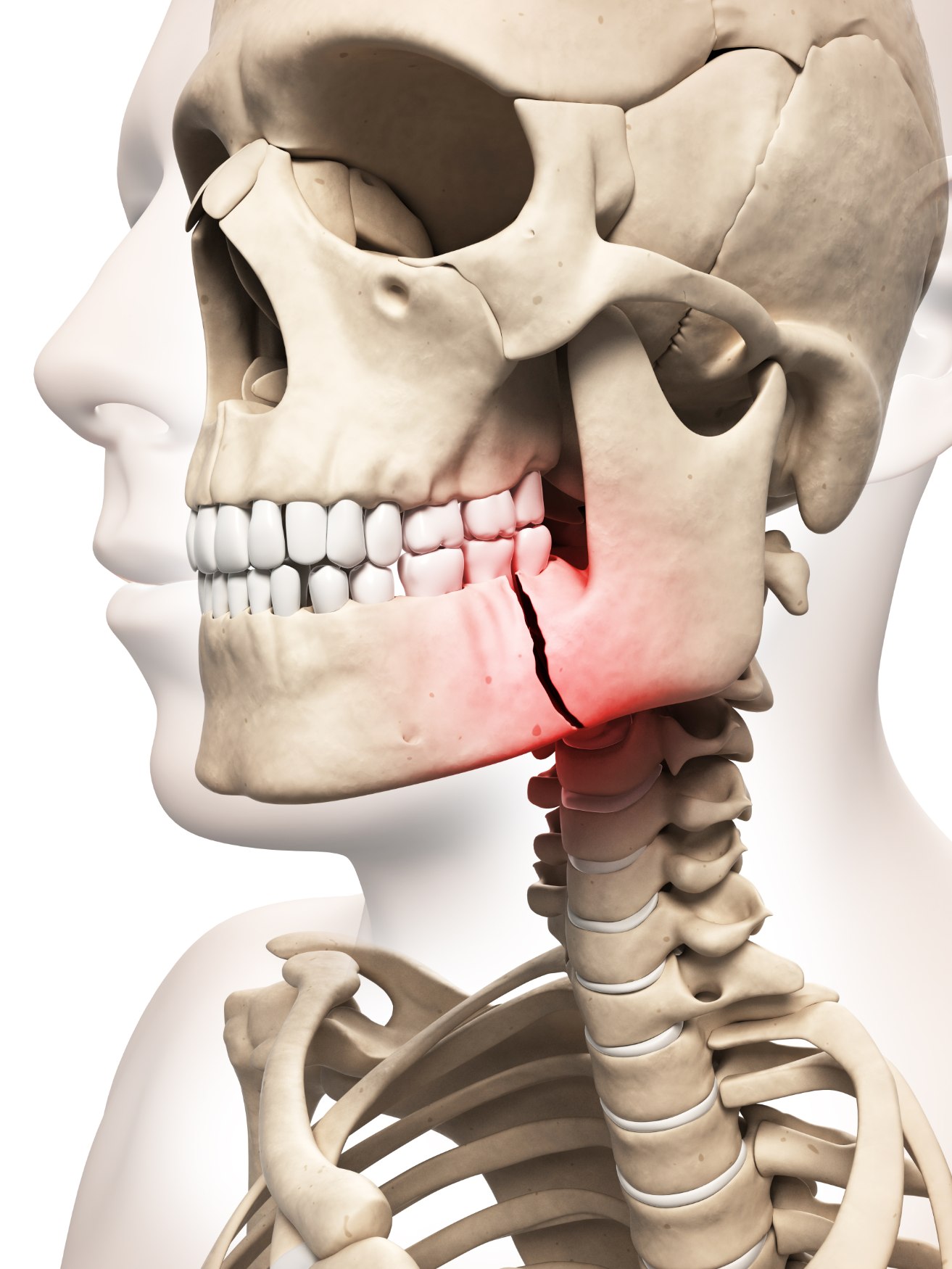
Facial fractures are unique in a way – if the bones are not aligned and fixed correctly, they usually lead to the deranged bite or a malocclusion. Depending on the location of the break or fracture, and the severity of the injury, your surgeon may offer a number of different treatment options. Duration of treatment can differ significantly depending on the individual condition, and in cases where there is only a minor fracture, effective management of the injury is still essential. Some jaw fractures have slight or no displacement, and the individual may look uninjured; however, the complaint of a subtle malocclusion leads their dentist to suspect a fracture.
In addition, an untreated jaw fracture can also result in lifelong facial deformity, which becomes very difficult to treat at a later stage. Our specialists use the latest diagnostic modalities like CBCT, CT scan, and Panorex scan to identify all the fractures and formulate a suitable treatment plan. They also use state-of-the-art 3D printing technology for the reconstruction of secondary traumatic defects, which are not susceptible to traditional treatment options.
In addition, an untreated jaw fracture can also result in lifelong facial deformity, which becomes very difficult to treat at a later stage. Our specialists use the latest diagnostic modalities like CBCT, CT scan, and Panorex scan to identify all the fractures and formulate a suitable treatment plan. They also use state-of-the-art 3D printing technology for the reconstruction of secondary traumatic defects, which are not susceptible to traditional treatment options.
Procedure
Sometimes, elastic bands may be used to guide the jaw into position following surgery. These are attached using small temporary metal wires or braces. Your procedure will be discussed with you in full during your consultation. Even the fractured zygomaticomaxillary complex can be managed and often treated from an intraoral approach, anatomically reduced and fixated in the presence of gross facial oedema. The days of the ‘Gillies’ lift technique are being replaced by the intra-oral buttress or other methods with rigid fixation. These facial fractures are often considered a simple treatment for Oral and Maxillofacial surgeons, as it is simply a matter of putting the puzzle pieces back together.